On this interview, NewsMedical talks to Jason Otterstrom in regards to the position of AI-based Athena Zebrafish software program in exploring Zebrafish imaging outcomes and its significance for bio-medical analysis utilizing Zebrafish fashions. is now obtainable for obtain with a versatile pay-per-use license.
How can high-content imaging of Zebrafish revolutionize the bio-medical analysis discipline?
Zebrafish (Danio rerio) are a promising emergent mannequin system for biomedical analysis with the potential to allow quick, excessive throughput research of human illness in dwelling vertebrates.
People and zebrafish have a lot of the similar organs, share 70% of the identical genes, and 84% of the genes related to human illness have zebrafish counterparts.
Zebrafish embryos are clear and will be manipulated on the single-cell stage, then shortly develop into larvae in a matter of days. This physiological growth will be imaged with a microscope utilizing 1000’s of particular person fish to instantly visualize the impact of therapies on organ techniques, growth and physiology.
No different well-characterized organic mannequin system is presently capable of provide these benefits.

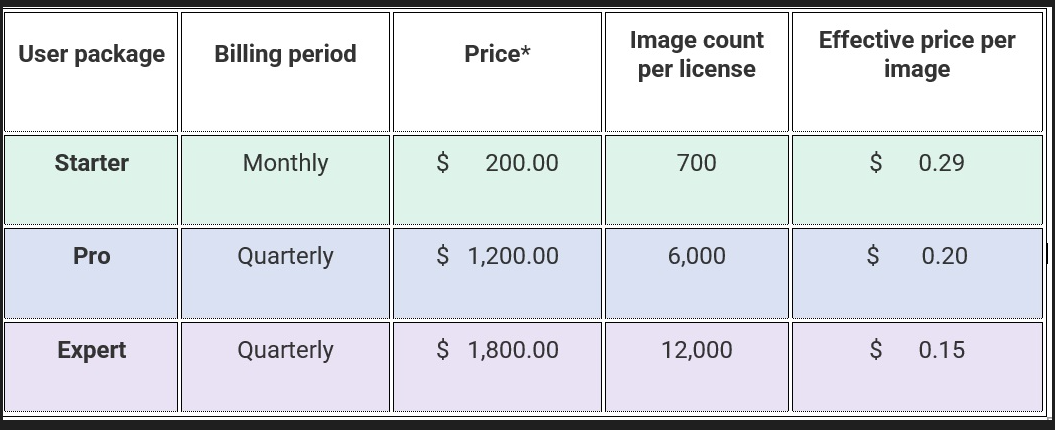
Dr. Otterstrom: “We provide three inexpensive license packages for various throughput wants. With an efficient worth between $0.15 and $0.29 USD per picture analyzed, any Zebrafish lab or researcher can use Athena software program to boost their evaluation workflows.” Picture Credit score: IDEA Bio-Medical Ltd.
How are photographs of zebrafish used, and why are they vital for researchers?
Microscopy photographs of zebrafish are analyzed to extract numerical or quantitative details about the embryos and larvae. The animal’s clear physique makes a lot of the fish’s inner anatomy observable, even with easy bright-field imaging.
Genetic manipulation permits the insertion of fluorescent proteins into the dwell zebrafish for tissue-specific imaging and evaluation.
Life science researchers then analyze photographs of zebrafish to review and measure organic and physiological modifications occurring in several anatomical areas inside a dwelling fish. For instance, organ growth and mobile proliferation or dying in relation to pharmacokinetics, toxicity, most cancers, and genetic modifications will be instantly noticed and measured.
How can AI assist in these investigations?
Analyzing photographs of zebrafish is a time-consuming bottleneck in screening research. Whereas the photographs could also be simply interpretable by a human, standard picture evaluation algorithms don’t readily detect the fish or its anatomy. As an alternative, measurements are carried out manually utilizing single photographs.
IDEA Bio-Medical discovered of this problem from our purchasers, so we created a novel deep-learning AI (synthetic intelligence) algorithm to routinely analyze their Zebrafish photographs for them.
The AI coaching course of is complicated and error-prone, requiring substantial effort and time that our biology-focused purchasers couldn’t do themselves. The AI now we have developed can now robustly establish objects just like the zebrafish contour and its inner anatomy which are clearly seen to us people.
We now need to make this software obtainable to all zebrafish researchers, regardless of which microscope they use.
IDEA’s Zebrafish evaluation software program was developed with our purchasers, for our purchasers.
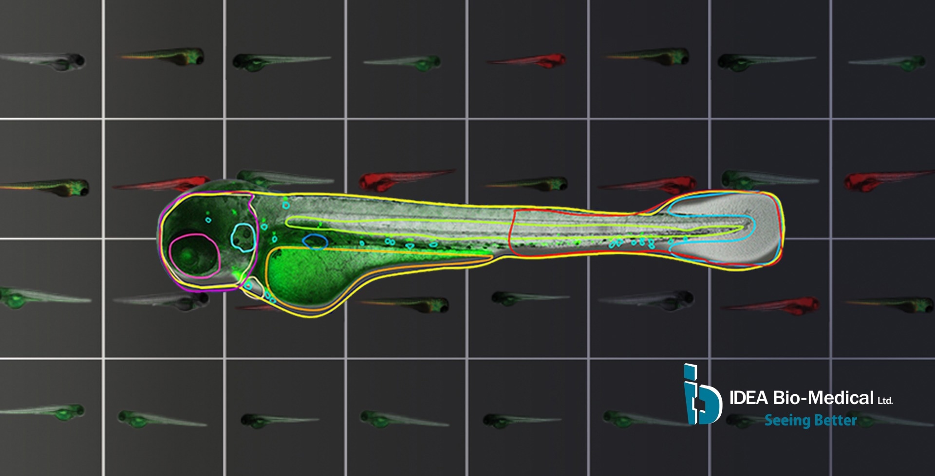
Athena software program permits parameter-free Zebrafish evaluation utilizing easy bright-field photographs, extracting the fish contour and far of its inner anatomy (yolk sac, eye, notocord and extra), together with physique areas of the top, trunk, and tail. Picture Credit score: IDEA Bio-Medical Ltd.
Why do we’d like details about Zebrafish anatomy to interpret fluorescence alerts?
Many processes investigated utilizing zebrafish happen or start in particular organs or areas inside the physique. They are often marked with fluorescence labeling to picture the tissue or cells of curiosity. Nevertheless, these have to be distinguished from undesired alerts and autofluorescence.
For instance, within the zebrafish larva, hematopoietic stem cells (HSC) originate in a particular space inside the tail – in any other case referred to as the caudal hematopoietic tissue (CHT). It’s analogous to the mammalian fetal liver and related to leukemia in people. Research trying on the proliferation of GFP-labeled HSC must focus particularly on the tail and ignore these fluorescent alerts current elsewhere, corresponding to circulating cells and yolk sac autofluorescence.
is presently the one software program obtainable that allows high-content screening on this context. It permits automated cell counts completely inside particular components of the fish to maximise each throughput and information extracted.
It contrasts widespread approaches, corresponding to easy depth thresholding, which ignores the seen anatomy. As an alternative, all fluorescence is gathered collectively, regardless of its origin, producing noisy information. Handbook choice of the tail would enhance high quality however severely restrict achievable throughput.
Athena is a first-of-its-kind automated picture evaluation software program devoted to zebrafish microscopy. Picture Credit score: IDEA Bio-Medical Ltd.
What’s Athena picture evaluation software program, and the way can it’s used?
is a stand-alone software program developed and provided by IDEA Bio-Medical to empower all zebrafish researchers. It allows true high-content imaging in zebrafish for the primary time.
Athena permits parameter-free zebrafish evaluation utilizing easy bright-field photographs. It routinely detects zebrafish embryos and larvae as much as 5 days outdated (dpf), extracting the fish contour and far of its inner anatomy (yolk sac, eye, notocord, and extra), together with physique areas of the top, trunk, and tail.
For every of those objects, the software program measures the morphology (space, size, and form) and might detect fluorescence in related shade channels. Each fluorescence depth and spot/construction detection inside particular anatomy are supported.
The software program is suited to a broad vary of researchers and accepts a number of picture format varieties output from almost all microscope producers.
Click on right here for a trial of Athena Zebrafish software program, obtainable for obtain from the IDEA Bio-Medical web site.
What benefits does this methodology have over different strategies?
Athena is the one automated software program devoted to zebrafish microscopy, providing a number of benefits:
- Automation: It permits experimental scale-up whereas eliminating subjective human bias or error and allowing direct comparability of outcomes between totally different labs.
- Intuitive and user-friendly interface: Customers can shortly and simply adapt Athena for his or her analysis.
- Versatile and versatile AI: Broadly relevant for a lot of varieties of screens involving zebrafish, as described by Athena prospects on the .
- Buyer assist with customized new consumer onboarding to make sure the success of our prospects.
What’s the Versatile Pay-Per-Use license you might be providing, and what are its benefits?
IDEA Bio-Medical acknowledges that annual software program licenses will be costly, particularly if photographs usually are not persistently analyzed all year long. Therefore, we provide Athena on a pay-per-use foundation in order that zebrafish researchers can adapt their license to investigate photographs after they want it.
We provide three inexpensive packages for various throughput wants. With an efficient worth between $0.15 and $0.29 USD per picture, any Zebrafish lab or researcher can use Athena and improve their evaluation workflows.
Licenses will be bought month-to-month or quarterly, based on want. This flexibility permits labs to regulate their licenses to suit their altering initiatives, college students, grant submissions and different dynamics of educational analysis.
Supply: IDEA Bio-Medical Ltd.
What assist does IDEA Bio-Medical present to Athena software program customers?
Analyzing microscopy photographs is complicated, requiring ability and expertise. Together with an intuitive software program interface, we provide customized buyer assist to allow any consumer of any ability degree to turn into unbiased and productive with Athena.
We at IDEA Bio-Medical take delight in working carefully with our purchasers and their distinctive wants. New customers can obtain a one-on-one onboarding session with a professional product specialist to stroll by way of the software program utilizing their very own information and guarantee a easy, constructive first expertise.
Extra Athena assist can be obtainable by reaching out to IDEA Bio-Medical instantly.
What’s the future route of your Athena software program, and who would profit from utilizing it?
Athena is a wealthy application-based picture evaluation software program for simple and fast evaluation of microscope photographs. The complete Athena package deal is presently built-in into the , IDEA Bio-Medical’s flagship excessive content material screening (HCS) microscope.
Sooner or later, the complete Athena software program, related for microscopy photographs of 2D and 3D cell tradition, will likely be provided as a stand-alone platform, permitting all biology researchers to carry out picture evaluation on the push of a button.
About Dr. Otterstrom
Dr. Otterstrom obtained a Bachelor of Arts in Utilized Physics on the College of Utah. As a Ph.D. scholar at Harvard College, he studied the biophysics of membrane fusion as mediated by influenza virus hemagglutinin protein utilizing single-molecule microscopy strategies. He went on to acquire a Marie Curie fellowship to make the most of super-resolution imaging to review chromatin fine-packing buildings on the Institute of Photonic Sciences (ICFO) close to Barcelona, Spain.
As an utility scientist for IDEA Bio-Medical, he helps purchasers with adapting their various experiments and assays to be carried out within the context of automated microscopy on the corporate’s flagship product, the WiScan Hermes.
About IDEA Bio-Medical Ltd.
was based in 2007 by way of a partnership between YEDA (the Weizmann Institute’s commercialization arm) and IDEA Machine Growth (an innovation hub).
The corporate focuses on automated imaging techniques and picture evaluation software program, providing a broad vary of organic purposes based mostly on the corporate’s distinctive algorithms library.
The system incorporates essentially the most superior applied sciences presently obtainable within the machine imaginative and prescient discipline, built-in with engineering methodologies of excessive reliability and high quality on the degree of semi-conductors and digital printing industries, that are the specialty of the mom firm,